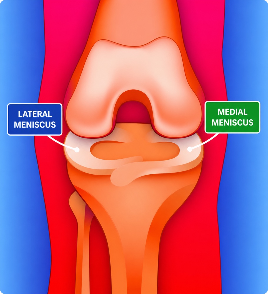
Anatomy and Function
Each knee contains two menisci: the medial meniscus on the inner side of the knee and the lateral meniscus on the outer side. They sit between the femur and tibia and help distribute force across the knee.
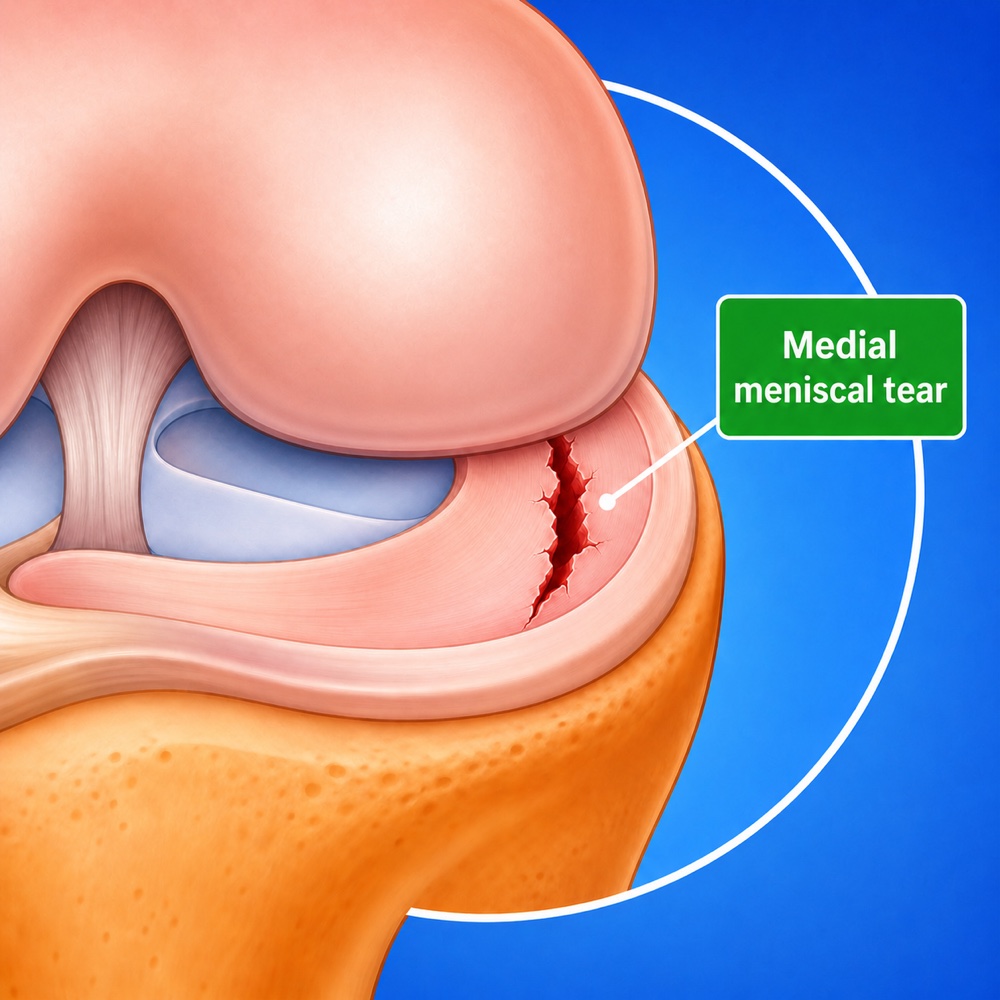
The outer third of the meniscus has a better blood supply and is often called the red zone. Tears in this region have greater healing potential. The inner portion has limited blood supply and is less likely to heal spontaneously or after repair.
The menisci protect articular cartilage by increasing contact area and decreasing peak joint stresses. Loss of meniscal tissue can increase contact pressure and may contribute to progression of degenerative change over time.